METHODS
METHODS
MAKING THE DSSCs
Figure 1 Drying Paste

The cells were created by finding the conductive side of a piece of conductive glass. They were then cut in to smaller pieces of around 1/2” by 1” and taped around the edge with scotch tape leaving a smaller area of space as seen in Figure 1, this is so that there was consistency in the size and the amount of paste in each cell. The paste as seen in Figure 1 was made when a beaker was taken and both TiO2 powder and concentrated acetic were stirred together, with a stir rod, until it had the consistency of paint. The paste itself was spread when the stir rod was used to scoop some paste up and evenly spread on the exposed conductive glass, then allowed to dry for 10 minutes.
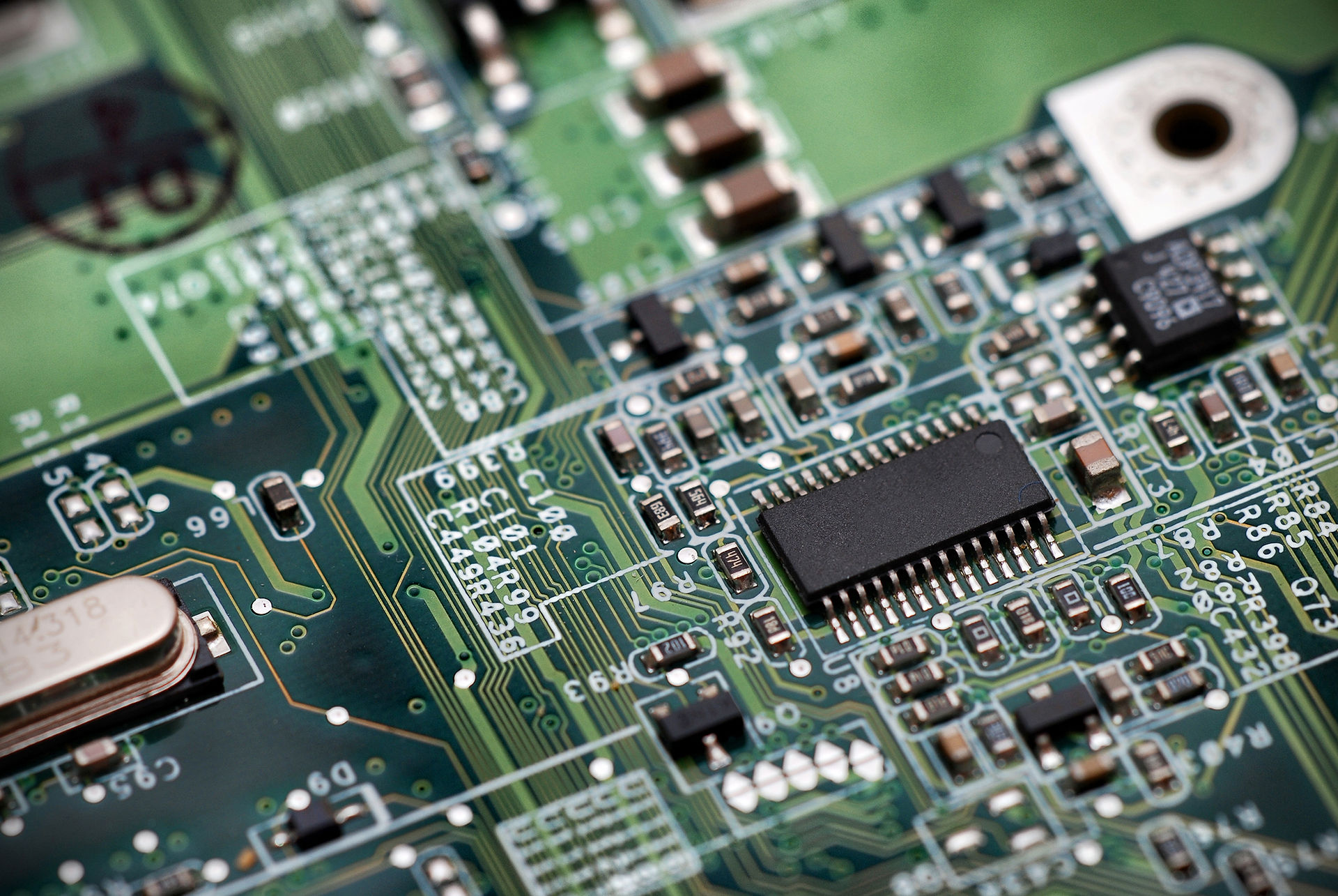
After the 10 minutes was over the cells’ tape were taken off and then they were placed on a hot plate at > 270 °C and allowed to heat up for 10 minutes and undergo sintering, as see in Figure 2, which allows the paste to become more porous and allow the dye to absorb better, this was observed by the paste turning brown for a few seconds then back to white once again, once the 10 minutes were over the cells were taken off the heat and allowed to cool down at room temperature.

Figure 2.2 Phase Transition/Sintering
Figure 2 Phase Transition/Sintering
Figure 3 Phase Transition/Sintering

Figure 4 Squished Berry Dye
Figure 3 Phase Transition/Sintering
Figure 3 Berry Reduction Dye
The berry reduction dye was created when another beaker was taken, and 5 black berries were put in a small amount of water. Then the beaker was put on to the hotplate and the berries were slowly broken down with a spoon to let the dye out of the berries, once the water was completely darkened as seen in Figure 3, the dye was ready to be used.
The squished berry dye was created when a Ziploc bag was taken and 4 blackberries were put inside and squished until only seeds, dye, and fruit skin could be seen, as seen in Figure 4. Then the dye and the fruit skin and seeds were separated from each other so that the dye could be easily accessed

Figure 5 Dyeing the Cells
Once both dyes were prepped two cells were designated to be dyed with the reduction and the other two were dyed with the squished berries. The dye was transported with a pipette and spread all over the TiO2 paste, as seen in Figure 5 so that all of it could absorb the dye for consistent current generation. Then the dyes were allowed to absorb at room temperature for 10 minutes.
Once the 10 minutes were over the cells were then washed with deionized water and isopropyl alcohol so that no excess dye would stay on the cell. The cells then were dabbed with KimWipesTM very gently, so that no dye was wiped off, until they were completely dry so that only the dyed TiO2 paste was present, on the cell, as seen in Figure 6

Figure 6 Cell Comparison


Figure 7 Soot Application
The top of the cell was made when another ½” x 1” piece of conductive glass was taken and put over a candle flame to allow a layer of soot to form, as seen in Figure 7. This layer of soot was so the electrolyte and the top of the cell could conduct better. Once the soot layer was added the glass was allowed to cool down enough so it could be handled with hands.
Once cooled down the dyed piece of glass and the soot piece of glass were connected using small binder clips, as seen in Figure 8. Once the cell was clipped together a potassium iodide electrolyte was added so that there would be electrons for the light to interact with and free for the dye to capture. A drop of electrolyte was added to the inside of the cell and allowed to soak over the dyed paste by alternatively clamping and un-clamping the binder clips, giving the yellow hue as seen in Figure 8. This was done to both the reduction cell and the squished berry cell. Once that was done the was ready test.
Figure 8 Phase Transition/Sintering

Figure 8 Completed Cell
TESTING THE CELLS
The cell was tested by using two multimeters in a loop so that the cell’s current and voltage could be measured, as seen in Figure 9. The multimeters were hooked up to a DC Power Supply, as seen in Figure 11, so that a voltage sweep could be performed.

Figure 9 Voltage and Current Multimeters

Figure 10 Cell Hookup
The cell was connected using alligator clips, the black wire was connected to the dyed part of the cell and the red wire was connected to the blackened part of the cell, as seen in Figure 8.
The cell was first tested at .01 V and, if the cell worked, there would be a negative current, this means that the cell is pushing more current, in the opposite direction, than the DC Power supply, which was set at 1/3 of its current knob. This was so that the cell was actually producing statistically significant current. From .01 V all the cells were tested in increments of .05 V all the way to .4V. Once all the data points were recorded, they were plotted on a graph in Excel.

Figure 11 DC Power Supply